Dive Brief:
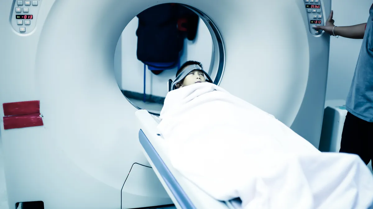
- The National Heart, Lung and Blood Institute of the National Institutes of Health has awarded a three-year $1.1 million grant to researchers at Case Western University School of Medicine to study a new imaging approach for diagnosing peripheral arterial disease (PAD) that does not involve the use of contrast dyes.
- The researchers are looking to develop techniques that avoid commonly used gadolinium-based contrast agents, which are a particular problem for patients with chronic kidney disease.
- The techniques to be studied are expected to be useful for other medical procedures as well, such as venography for blood flow, vessel wall imaging used in stroke diagnosis, and perfusion imaging used to see blood flow in tissues.
Dive Insight:
Gadolinium-based contrast agents injected into the body to improve the quality of of magnetic resonance imaging (MRI) scans have been the subject of some debate in recent years due to concerns about longer-term retention of the substances in organs. A 2010 safety announcement from the FDA warned of an association with nephrogenic systemic fibrosis in patients with kidney dysfunction.
Mayo Clinic researchers in 2015 found evidence of gadolinium deposits in brain tissue samples from patients who had undergone MRI exams and donated their bodies to medical research.
The FDA evaluated the risk of brain deposits with repeated use of gadolinium-based contrast agents for MRI and determined in 2017 that the metal is retained in the brain, bones and skin. The agency did not find adverse health effects related to gadolinium in the brain. Still, the FDA in December announced a new class warning about gadolinium retention to appear in the labeling for gadolinium-based contrast agents used in MRI.
The Case Western Reserve researchers noted that chronic kidney disease is becoming more common in the United States due to rising rates of diabetes and hypertension. At the same time, more than 200 million people worldwide suffer from PAD, a potentially serious circulatory problem in the legs.
Sanjay Rajagopalan, director of the Cardiovascular Research Institute at Case Western Reserve University School of Medicine, and his colleagues will focus on a technique for non-contrast-enhanced imaging that magnetically labels flowing blood and generates paired images of the tissue to allow clear visibility of blood vessels without the need for contrast dye. The clinical studies will include comparisons to contrast-enhanced images.
The researchers also will look at the feasibility of high-rate scan acceleration using compressed sensing combined with parallel imaging.


