Dive Brief:
-
The NIH has awarded researchers $3.8 million to test a way of diagnosing suicidal thinking from images of the brain.
-
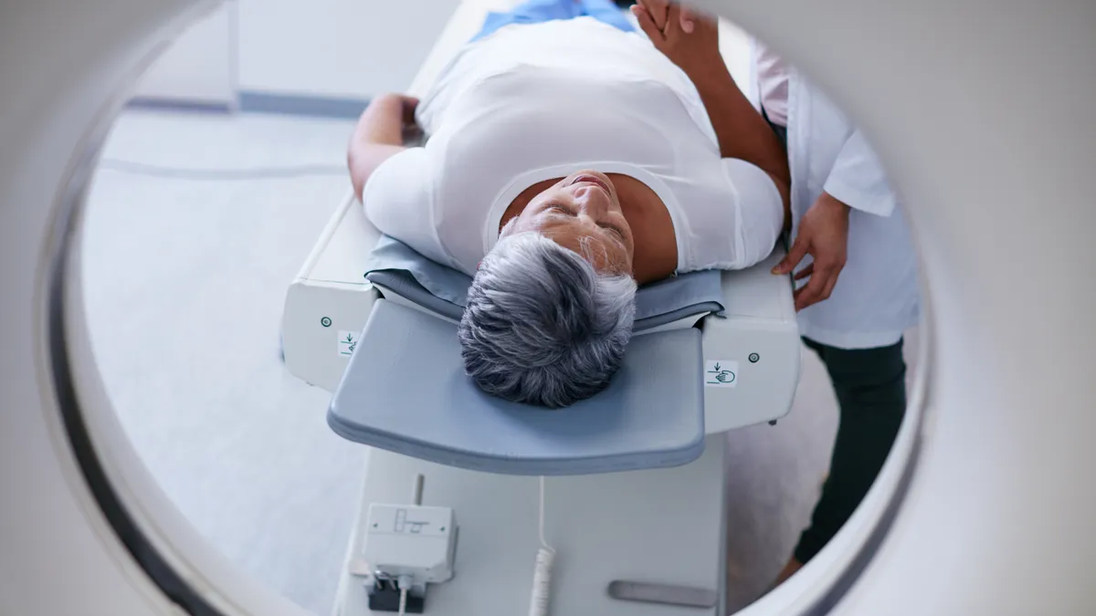
Researchers will use functional magnetic resonance imaging (fMRI) to analyze how the brain activity of young adults changes when they think about words related to suicide.
-
The latest study builds on a paper published last year, which suggested fMRI can predict whether someone is suicidal, but left scope for doubt about the replicability of its findings.
Dive Insight:
Researchers have used fMRI to analyze people’s responses to words for years. These studies have people view images or think of words while lying in MRI scanners and monitor how their brain’s light up, providing evidence of correlations between neural pathways and certain ideas or objects. Last year, a team applied this idea to suicide and published their findings in Nature Human Behaviour.
“We were previously able to obtain consistent neural signatures to determine whether someone was thinking about objects like a banana or a hammer by examining their fMRI brain activation patterns. But now we are able to tell whether someone is thinking about 'trouble' or 'death' in an unusual way. The alterations in the signatures of these concepts are the 'neurocognitive thought markers' that our machine learning program looks for,” Carnegie Mellon University's Marcel Just said in a statement.
As many people hide their suicidal intentions, it can be hard for healthcare professionals to identify who is at risk. The 34-person study run by Just and collaborator David Brent claimed to show a way to look inside people’s brains and objectively state whether they were suicidal. Just and Brent have now received NIH funding to run a larger study to validate the findings.
Carnegie Mellon is touting the study as a precursor to the approach being used in clinical practice, where it will enable healthcare professionals to detect and monitor suicidal risk, understand altered patterns of thinking and feeling in their patients and develop treatments tailored to these alterations.
However, there are reasons to doubt whether the approach will enter clinical practice, even if the larger study replicates the findings of the original trial. MRI is expensive and requires patients to stay still in a stressful environment for long periods of time. In many healthcare centers, demand for the imaging equipment exceeds capacity, leading to long wait times for patients needing MRIs.
It is also questionable whether the test will be accurate enough. In the original trial, the test had a specificity of 0.94 and a sensitivity of 0.88. While that means the test is accurate most of the time, it could still result in a enough false positives and false negatives to render it unhelpful at scale.
These and other doubts will hang over the approach until a more comprehensive assessment of its effectiveness is complete. With funding in place, Just and Brent are a step closer to performing such an assessment.


