Dive Brief:
- Microsoft has partnered with pathology software provider Paige to develop the “world’s largest image-based artificial intelligence (AI) models” in oncology.
- Paige, the developer of an AI-based pathology product cleared by the Food and Drug Administration, has already developed a large foundation model using more than 1 billion image types. Microsoft will support development of a new model that is “orders-of-magnitude larger” than any in use today.
- Using Microsoft’s supercomputing and cloud-computing infrastructure, Paige aims to build an AI model that can generate new insights into the pathology of cancer to improve care.
Dive Insight:
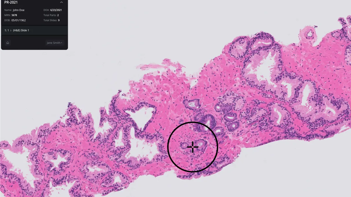
Paige has scaled up its models since launching in New York’s Memorial Sloan Kettering Cancer Center. To develop its first authorized product, Paige Prostate Detect, the company trained its AI on 60,000 slide images and used a further 40,000 slides to validate its ability to detect potentially cancerous areas. As it has expanded into other cancers, Paige has built a model based on more than 500,000 pathology slides.
Those slides, which feature more than 1 billion images of multiple cancer types, are the start of a bigger plan. For the next stage of the project, Paige is incorporating up to 4 million digitized microscopy slides.
Microsoft is supporting the jump in scale. Paige has identified Microsoft’s supercomputing infrastructure as a resource that can enable it to train its model at scale, and ultimately aims to use the tech company’s Azure cloud platform to make the AI capabilities available to hospitals and laboratories globally.
“By combining Microsoft’s world-class research and cloud infrastructure with Paige’s deep expertise and large-scale data, we are creating new AI models that will enable unprecedented insights into the pathology of cancer,” Desney Tan, vice president and managing director, Microsoft Health Futures, said in a statement.
Paige sees the partnership as a way to both increase accuracy and unlock new capabilities. Achieving that goal could give pathologists additional support as they interpret images to determine the condition of the patient.

